Ferroptosis and Reagent Selection Guide
What is Ferroptosis?
"Ferroptosis" was coined by Stockwell et al. at Columbia University in 2012 and described as a form of iron-dependent cell death. * It was reported to be a form of programmed cell death by the Nomenclature Committee on Cell Death (NCCD) in 2018.
Ferroptosis is a form of programmed cell death that is caused by iron ion-dependent accumulation of lipid peroxides. Ferroptosis has been shown to follow a different cell death pathway from apoptosis and thus is attracting attention as a new target for cancer therapy. It has also been found to be associated with various diseases, such as neurodegenerative diseases, cerebral apoplexy, and hepatitis (NASH).
*S. J. Dixon, B. R. Stockwell et al., Ferroptosis: An iron-dependent form of nonapoptotic cell death., Cell, 2012, 149(5), 1060.
How Does Ferroptosis Cause Cell Death?
|
|
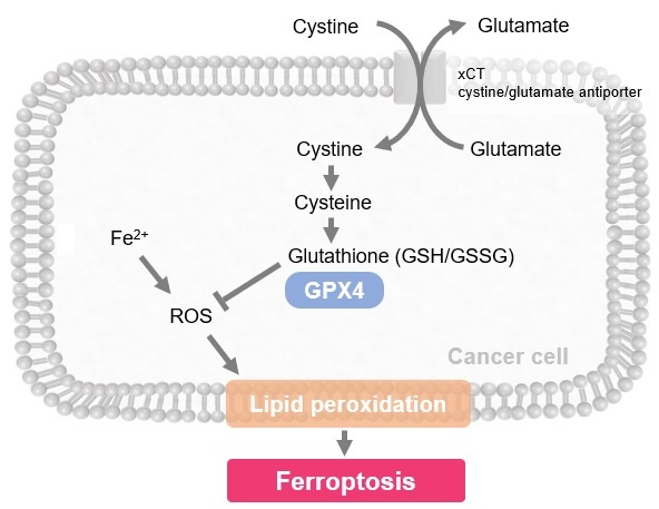
Ferroptosis is characterized by the accumulation of lipid peroxides. Lipid peroxides are formed from oxidation of polyunsaturated fatty acids (PUFA) in membrane phospholipids, with iron suggested to be involved. Intracellular glutathione peroxidase 4 (GPX4) uses reduced glutathione (GSH), an antioxidant, to reduce lipid peroxides generated by reactive oxygen species (ROS).*
However, when lipid peroxides accumulate due to GPX4 disruption or GSH depletion, ferroptosis is triggered.
* Stockwell et al, a leading researcher in the field of ferroptosis, summarized inhibitors, inducers, and detection indicators of ferroptosis in the following review, in which Dojindo’s Liperfluo is introduced for detection of lipid peroxides.
B. R. Stockwell, et al., "Ferroptosis: A Regulated Cell Death Nexus Linking Metabolism, Redox Biology, and Disease.", Cell, 2017, 171, 273.
|
Induction of Ferroptosis by Erastin?
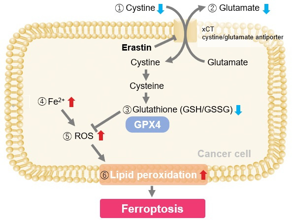
Erastin is a known inducer of ferroptosis. By inhibiting the cystine transporter (xCT), erastin inhibits the uptake of cystine. Cystine is the raw material for GSH. Therefore, Erastin ultimately decreases the amount of GSH. Decreased GSH then results in lipid peroxide accumulation and induction of ferroptosis.
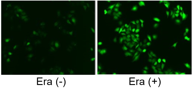
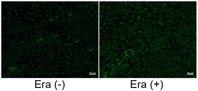
The following experimental examples show changes in each aforementioned index as a consequence of erastin stimulation. Measurements are made using Dojindo reagents.
<Experimental example>
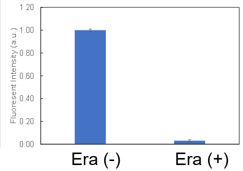
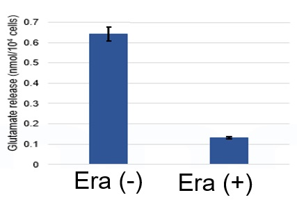
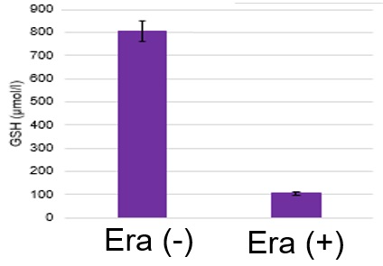
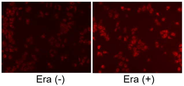
Using erastin-treated A549 cells, we measured intracellular Fe
2+, ROS, lipid peroxide, glutathione, glutamate release into the extracellular space, and cystine uptake. As a result, inhibition of xCT by elastin was observed and also the release of glutamate and uptake of cystine were decreased. Furthermore, elastin treatment decreased intracellular glutathione while it increased intracellular Fe
2+ , ROS, and lipid peroxides.
Research on Related Diseases
Nonalcoholic steatohepatitis (NASH)
|
Suppression of hepatitis via ferroptosis
In a study involving the livers of NASH model mice, it was confirmed that necrosis precedes apoptosis in the development of fatty liver. Further experiments showed that ferroptosis is involved within necrosis as a trigger for steatohepatitis and that inhibition of ferroptosis almost completely suppressed the onset of hepatitis.
Minoru Tanaka, et al., "Hepatic ferroptosis plays an important role as the trigger for initiating inflammation in nonalcoholic steatohepatitis", Cell Death & Disease, 2019, 10, 449.
Related article: changes in intracellular markers associated with NASH
The article summarizes reports on changes in each indicator of metabolic states and cellular senescence using the NASH model.
(Click on the "NASH" tab in the link)
Experimental example: measurement of intracellular metabolism in NASH model tissue
Measurement of ATP, a-KG, and NAD levels in liver tissue of high-fat diet-treated type 1 diabetic model mice. (Please refer to each product's website for more information, “Experimental Example: Change in Metabolism in Liver Tissue of NASH-Induced Mouse”)
|
|
|
Neurodegenerative disease
|
Confirmation of the link between lysosomal disorders and ferroptosis
In experiments using human neurons, it is reported that knockdown of the lysosomal protein prosaposin induces formation of lipofuscin, a hallmark of aging. This process involves the iron-catalyzed generation of reactive oxygen species, leading to induction of ferroptosis.
Martin Kampmann, et al., "Genome-wide CRISPRi/a screens in human neurons link lysosomal failure to ferroptosis", Nature Neuroscience, 2021, 24, 1020
|
|
|
Cancer
|
Regulation of cancer immunity via ferroptosis
CD8+ T cells activated by immunotherapy were found to confer an anti-tumor effect by promoting lipid peroxidation and inducing ferroptosis. The mechanism of immunotherapy-induced inhibition of cystine uptake and promotion of lipid peroxidation in tumor cells is discussed.
Weiping Zou, et al, "CD8+ T cells regulate tumour ferroptosis during cancer immunotherapy", Nature, 2019, 569, 270
|
|
|
Ferroptosis
|
Ferroptosis - a newly identified, iron-dependent form of programmed cell death
A summary of the current progress in studying ferroptosis, as well as its potential applications in the fields of biology and medicine.
Fudi Wang, et al., "Ferroptosis: Beauty or the Beast", Dojin News, 2021, 178, 1
|
|
Ferroptosis-Related Reagent Selection Guide
Lipid Peroxide and Iron (Fe2+) Detection Reagents
Name
|
Liperfluo
|
MitoPeDPP
|
Mito-FerroGreen
|
FerroOrange
|
Target
|
Lipid Peroxidation
|
Lipid Peroxidation
|
Ferrous Ion(Fe2+)
|
Ferrous Ion(Fe2+)
|
Localization
|
Intracellular
|
Mitochondria
|
Mitochondria
|
Intracellular
|
Detection (Fluorescence:Ex/Em)
|
Fluorescence (524 nm/535 nm)
|
Fluorescence (452 nm/470 nm)
|
Fluorescence (505 nm/580 nm)
|
Fluorescence (543 nm/580 nm)
|
Instrument
|
Fluorescence Microscope, FCM
|
Fluorescence Microscope, FCM
|
Fluorescence Microscope, Microplate Reader
|
Fluorescence Microscope
|
Sample
|
Live Cell
|
Live Cell
|
Live Cell
|
Live Cell
|
Oxidative Stress- and Metabolism-Related Reagents and Kits
Name
|
ROS Assay Kit -Highly Sensitive DCFH-DA-
|
GSSG/GSH Quantification Kit
|
Glutamine Assay Kit-WST
|
Glutamate Assay Kit-WST
|
Target
|
ROS (Reactive oxygen species)
|
Glutathione (oxidized/reduced)
|
Glutamine
|
Glutamine
|
Localization
|
Intracellular
|
Intracellular
|
Intracellular/Extracellular
|
Intracellular/Extracellular
|
Detection (Fluorescence:Ex/Em)
|
Fluorescence (505 nm/525 nm)
|
Colorimetric:412 nm
|
Colorimetric:450 nm
|
Colorimetric:450 nm
|
Instrument
|
Fluorescence Microscope, FCM,
Microplate Reader
|
Microplate Reader
|
Microplate Reader
|
Microplate Reader
|
Sample
|
Live Cell
|
Cell, Tissue, Blood Plasma, Red Blood Cell
|
Cell, Cell Culture
|
Cell, Cell Culture
|
Related Scientific Information