Cellular Senescence Reagent Selection Guide

What is Cellular Senescence?

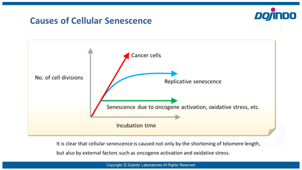
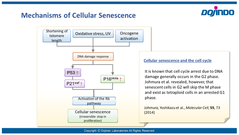
 Cellular senescence was reported by Hayflick in 1981. It was discovered when pulmonary fibroblasts slowed down their proliferation and eventually ended in cell death after cell passaging had been performed for more than 8 months. Subsequent studies have revealed that cellular senescence is caused not only by telomere length reduction, but also by external factors such as oncogene activation, oxidative stress, and DNA damage.
The induction and control mechanisms of cellular senescence – in which genetic and external factors are intricately involved – have yet to be fully elucidated. However, it has been suggested that the process is closely related to cancer and various age-related diseases, inspiring large amounts of active research into the topic. The development of drugs that eliminate senescent cells in the body (senolytic drugs) is also attracting the attention of researchers as a possible strategy to extend healthy life expectancy.

Assessing Cellular Senescence

 Cellular senescence is controlled by various factors such as cell type and physiological conditions, such as oxidative stress. None of the individual biomarkers that have been identified so far have been deemed to be specific to senescent cells. Therefore, it is desirable to determine and confirm cellular senescence using multiple indicators.
 Common detection indicators for assessing cellular senescence include features related to cell cycle progression (DNA synthesis, p16/p21 expression, etc.), features related to morphology (of the cell, nucleus, nucleolus, etc.), SA-ß-Gal activity, DNA damage, oxidative stress (ROS), telomere length, inflammatory cytokines (senescence-associated secretory phenotype (SASP)), and more.

Learn more about cellular senescence

“Recent Findings of Cellular Senescence Studies and Analysis Method”

Chapters:
0:00 What is Cellular Senescence ?
5:00 Senescence Studies and Drug discovery
12:30 Methods of Senescence Detection and Analysis

Research on Cellular Senescence

Biomarker of Cellular Senescence

University of Cambridge, Dr. Muñoz-Espín

The article illustrates how markers are used to determine cellular senescence, using charts and graphs. The paper proposes using cell cycle indicators, SA-ß-Gal, and nuclear features as primary markers of cellular senescence.

Estela González-Gualda, Andrew G Baker, Ljiljana Fruk, Daniel Muñoz-Espín, A guide to assessing cellular senescence in vitro and in vivoFEBS J., 2021, 288, 56.

Senolytic CAR T Cells: Targeting Senescence-Related Diseases

Memorial Sloan Kettering Cancer Center, Dr. Corina Amor

Ever since recent research has reported that the removal of senescent cells can prolong a healthy lifespan, the development of senolytic drugs has become popular.
Amor et al. performed a study involving the attempted removal of senescent cells via use of CAR T cells. The results show the successful removal of senescent cells and the reduction of liver fibrosis. This study implies that the use of CAR T cells may work not only as anticancer therapy but also as a potential therapy for other diseases.

Amor, C. et al., Senolytic CAR T cells reverse senescence-associated pathologiesNature., 2020, 583, 127–132.

Cellular Senescence, Aging and Age-Related Disorders

Division of Cancer Cell Biology, Institute of Medical Science, The University of Tokyo, Dr. Yoshikazu Johmura, Dr. Nakanishi Makoto

Recently, many of the mechanisms by which cellular senescence is induced and maintained have been uncovered. These same mechanisms also have implications in aging, age-related disorders, and carcinogenesis in vivo. The discovery of these connections has had a great impact on senolytics research.

Y. Johmura, et al., Senolysis by glutaminolysis inhibition ameliorates various age-associated disordersSCIENCE2021, 371(6526), 265-270.

Indicators Related to Cellular Senescence

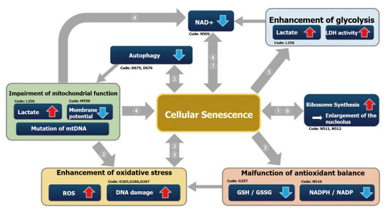
Correlation map of related indicators

 Research into apoptosis, necrosis, autophagy, and cellular senescence is very important for understanding the intracellular functions that control cell survival and death.
Recently, various fields have given particular attention to cellular senescence due to the recent discoveries of SASP (a known cancer-causing factor) and senescence-related phenomena in stem cell research.

 This correlation map shows the relationship between various intracellular indicators, resulting from cellular senescence. This information is based on currently available information. Please refer to the table with cited references below as reference for your experiments. The table lists the cell type, the method of senescence induction used, the senescence markers measured, and the variables affected by senescence in each reference for the map.

References cited ①-⑦ above

  Cell Senescence induction Senescence marker (s) Responding variable (s) Reference
IMR90
(Human pulmonary fibroblasts)
Several passages in culture SA-ß-Gal, p16, p21, Nucleosome hypertrophy Expression of SETD8↓,  H4K20me1↓, oxidative phosphorylation↑, ribosome synthesis↑ H. Tanaka, S. Takebayashi, A. Sakamoto, N. Saitoh, S. Hino and M. Nakao, “The SETD8/PR-Set7 Methyltransferase Functions as a Barrier to Prevent Senescence-Associated Metabolic Remodeling.”Cell Reports2017, 18(9), 2148.
Inhibition of SETD8
(Methyltransferase)
Oxidative phosphorylation↑,  ribosome synthesis↑
Senescent mouse satellite cell
eletal muscle progenitor cells)
SA-ß-Gal, p16 Autophagy activity↓, ROS↑, mitochondrial membrane potential L. Garcia-Prat, M. Martinez-Vicente and P. Munoz-Canoves, “Autophagy: a decisive process for stemness”Oncotarget2016, 7(11), 12286.
Atg7 knockout mouse
(Satellite cells)
Autophagy inhibition SA-ß-Gal, P15, p16, p21, γ-H2AX ROS↑, mitochondrial membrane potential
Rat fibroblast model of type 2 diabetes SA-ß-Gal, p21, p53, γ-H2AX NADP+/ NADPH↓(resistance to oxidative stress↓), NADPH oxidase↑(ROS↑) M. Bitar, S. Abdel-Halim and F. Al-Mulla, “Caveolin-1/PTRF upregulation constitutes a mechanism for mediating p53-induced cellular senescence: implications for evidence-based therapy of delayed wound healing in diabetes”Am J Physiol Endocrinol Metab.2013, 305(8), E951.
IMR90
(Human pulmonary fibroblasts)
Ethidium bromide (inhibition of mtDNA) + pyruvate deficiency SA-ß-Gal NAD+/NADH C. Wiley, M. Velarde, P. Lecot, A. Gerencser, E. Verdin, J. Campisi, et. al., “Mitochondrial Dysfunction Induces Senescence with a Distinct Secretory Phenotype”Cell Metab., 2016, 23(2), 303.
MDA-MB-231
(Human breast cancer cells)
X-ray irradiation + inhibition of cell cycle-related factor (securin) expression SA-ß-Gal Lactate↑, LDH activity↑, (glycolysis↑) E. Liao, Y. Hsu, Q. Chuah, Y. Lee, J. Hu, T. Huang, P-M Yang & S-J Chiu, “Radiation induces senescence and a bystander effect through metabolic alterations.”Cell Death Dis., 2014, 5, e1255.
MEF
(Mouse Embryonic Fibroblast)
Overexpression of oncogenes,several passages in culture, transcription factor overexpression(E2F1) SA-ß-Gal, p16, p21, Nucleosome hypertrophy Ribosome RNA↑, p53↑ K. Nishimura, T. Kumazawa, T. Kuroda, A. Murayama, J. Yanagisawa and K. Kimura, “Perturbation of Ribosome Biogenesis Drives Cells into Senescence through 5S RNP-Mediated p53 Activation”Cell Rep2015, 10(8), 1310.
Mouse tail fibroblast 2 months old, 22 months old, p16 knockout (22 months old) SA-ß-Gal, p14, p16 NAD+↓, SIRT3↓ M. J. Son, Y. Kwon, T. Son and Y. S. Cho, “Restoration of Mitochondrial NAD+ Levels Delays Stem Cell Senescence and Facilitates Reprogramming of Aged Somatic Cells”Stem Cells2016, 34(12), 2840.

Cell Cycle Arrest

 Irreversible cell cycle arrest is one of the phenomena that characterize cellular senescence. p16, p21, p53, and pRB (phosphorylated retinoblastoma protein) are known as representative protein markers. The activation/upregulation of these proteins are used as indicators of cellular senescence. These marker proteins are known to be tumor suppressors and regulate the cell cycle mainly through two pathways (p16Ink4a-RB and p53-p21CIP1).

‹Experimental Example›

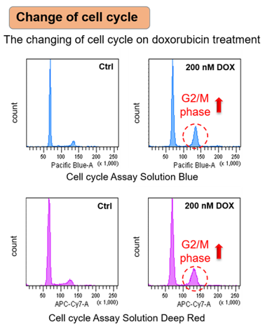
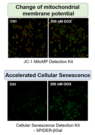
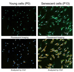
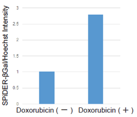
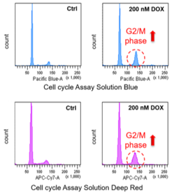
Doxorubicin (DOX) is known as an anticancer drug that acts in the G2/M phase of the cell cycle to arrest cell proliferation and induce cellular senescence (see the figure below in center). Below are the results of an experiment in which DOX was added to A549 cells. As a result, changes in SA-ß-Gal expression, cell cycle progression, and mitochondrial membrane potential were observed.

Changes in Intracellular Metabolism

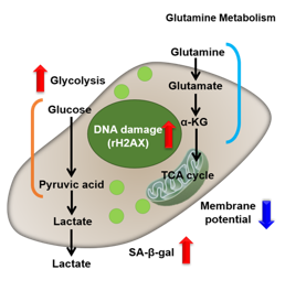
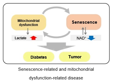
 It is well known that senescent cells maintain high metabolic activity despite their reduced proliferative capacity. In general, senescent cells show a decrease in NAD+, an increase in lactate efflux, and a decrease in AMP/ATP ratio. This is due to conversion from oxidative phosphorylation to aerobic glycolysis and mitochondrial dysfunction, in addition to activation of the glycolytic system.
Changes in intracellular metabolism are thus closely related to cellular senescence. Therefore, these changes in intracellular metabolism are very important – not only as indicators of cellular senescence, but also in clinical and basic research targeting cellular senescence.
Our webpage on intracellular metabolism provides maps focusing on senescence-associated changes in intracellular metabolism, such as SIRT1-related changes in NAD+ levels, and cells that have become senescent due to DNA damage.
(Please click on the “Senescence” tab in the link)

Lipid Accumulation (Lipotoxicity)

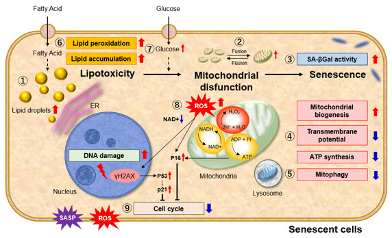
 Lipotoxicity, which is caused by excessive lipid accumulation in non-adipose tissue cells, is thought to be involved in cancer, diabetes, heart failure, and obesity. It has been shown that lipid accumulation in cells causes cellular senescence and mitochondrial dysfunction. The figure below shows the changes in various indices caused by excessive lipid accumulation.

For more information click here or image below

References

Clara, C. et al., “Mitochondria: Are they causal players in cellular senescence?”, Biochimica et Biophysica Acta – Bioenergetics, 2015, 1847(11), 1373-1379.

Huizhen, Z. et al., “Lipidomics reveals carnitine palmitoyltransferase 1C protects cancer cells from lipotoxicity and senescence”, Journal of Pharmaceutical Analysis, 2020.

Xiaojuan, H. et al., “Astrocyte Senescence and Alzheimer’s Disease: A Review”, Front. Aging Neurosci., 2020.

Borén, J. et al., “Apoptosis-induced mitochondrial dysfunction causes cytoplasmic lipid droplet formation”, Cell Death Differ, 2012, 19(9), 1561-1570.

Na, L. et al., “Aging and stress induced ß cell senescence and its implication in diabetes development”, Aging (Albany NY), 2019, 11(21), 9947–9959.

Cellular Senescence Reagent Selection Guide

Dojindo offers four types of kits and reagents that can be selected according to the evaluation method and purpose of cell senescence.

Product Cellular Senescence Detection Kit – SPiDER-ßGal Cellular Senescence Plate Assay Kit – SPiDER-ßGal Cell Cycle Assay Solution Deep Red / Blue Nucleolus Bright Green / Red
Detection Fluorescence Fluorescence Fluorescence Fluorescence
Wavelength
(Ex/Em)
Ex. 500 – 540 nm /
Em. 530 – 570 nm
Ex. 535 nm / Em. 580 nm Deep Red: Ex. 633-647 nm /
Em. 780/60 nm
Blue: Ex. 405-407 nm /
Em. 450/50 nm
Green: Ex. 513 nm /
Em. 538 nm
Red: Ex. 537 nm /
Em. 605 nm
Target SA-ß-gal activity SA-ß-gal activity Nucleus Changes in the nucleolus
Detection
Method
Imaging
Substrate: SPiDER-ßGal
Plate assay
Substrate: SPiDER-ßGal
Flow cytometry Imaging Detection of the nucleolus by RNA-staining reagent
Instrument Fluorescence microscope,
FCM
Fluorescence microplate reader FCM Fluorescence microscope
Sample Live cells, fixed cells
(Tissue: some examples from published articles using SG02)
Live cells
(lysis of live cells)
Live cells, fixed cells Fixed cells
Best for Those who have difficulty quantifying data or performing multiple staining with X-gal Those who process multiple samples
Those who are evaluating senescent cells for the first time Small size package (20 tests) is available
Those who wish to evaluate using indicators other than SA-ß-Gal Those who wish to evaluate using indicators other than SA-ß-Gal
Examples of reports using nucleolus as an indicator are available on the product page
data
Item# SG03 SG05 Deep Red: C548
Blue: C549
Green: N511
Red: N512

Product Classification

Product Classification