


-Cellstain- AO solution

Nucleus Dye
-
Product codeA430 -Cellstain- AO solution
-
CAS No.65-61-2(AO)
-
Chemical name3,6-Bis(dimethylamino)acridine, hydrOchloride, solution
-
MWC17H20ClN3=301.81
| Unit size | Price | Item Code |
|---|---|---|
| 1 ml | A430-10 |
Description
Product Description
Acridine orange (AO) forms a complex with double-stranded DNA to emit green fluorescence (Fig. 1). AO also forms a complex with singlestranded DNA or RNA to emit red fluorescence. One molecule of AO intercalates with three base pairs of double-stranded DNA and emits green fluorescence with the maximum wavelength at 526 nm (excitation 502 nm). One molecule of AO can also interact with one phosphate group of single-stranded DNA or RNA to form an aggregated, or stacked, structure that emits red fluorescence with the maximum wavelength at 650 nm (excitation 460 nm). Therefore, AO is utilized for the detection of both double-stranded DNA and single-stranded DNA or RNA. It enables simultaneous determination of DNA and RNA with argon laser excitation or flow cytometry.
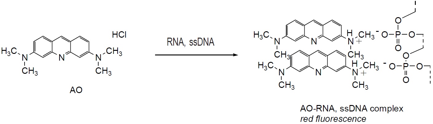
Fig. 1 Cell staining mechanism
Technical info
Staining Procedure
1.Prepare 10-50 μM AO solution with PBS or an appropriate buffer.a)
2.Add AO solution with 1/10 of the volume of cell culture medium to the cell culture.b)
3.Incubate the cell at 37ºC for 10-20 min.
4.Wash cells twice with PBS or an appropriate buffer.
5.Observe the cells under a fluorescence microscope with 500 nm excitation and 530 nm emission filters.
a) Since AO may be carcinogenic, extreme care is necessary during handling.
b) You may replace the culture medium with 1/10 concentration of AO buffer solution.
Data
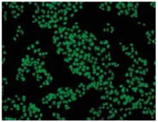
Fig. 2 Cell staining with AO Cell type: HeLa
References
1. I. W. Taylor, et al., An Evaluation of DNA Fluochromes, Staining Techniques, and Analysis for Flow Cytometry. I. Unperturebed Cellpopulations. J Histochem Cytochem. 1980;28:1224-1232.
2. N. Miyoshi, et al., Fluorescence Lifetime of Acridine Orange in Sodiu Dodecyl Sulfate Premicellar Solutions. Photochem Photobiol. 1988;47:685-688.
3. A. K. El-Naggar, et al., Single- and Double-stranded RNA Measurements by Flow Cytometry in Solid Neoplasms. Cytometry. 1991;12:330-335. 4. Y. Miyakoshi, et al., The Frequencies of Micronuclei Induced by Cisplatin in Newborn Rat Astrocytes Are Increased by 50-Hz, 7.5- and 10-mT Electromagnetic Fields. Environ Health and Prev Med. 2005;10:138-143.
Handling and storage condition
| Appearance: | Yellow to orange liquid |
|---|---|
| Dye content: | To pass test |
| -20°C, Protect from light |
Related products
The following related products are also used in research related to this product.


