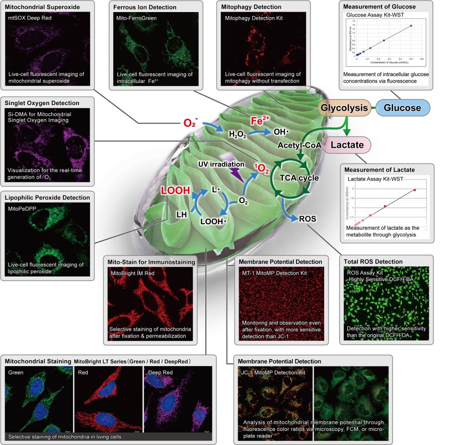
Mitochondria Research
Mitochondria, which have important functions in cells, are not only a place for ATP production by oxidative phosphorylation, but their activity and dysfunction are closely related to cancer, senescence, and neurodegenerative diseases such as Alzheimer's disease and Parkinson's disease. Therefore, understanding the state of mitochondria is particularly important.
Mitophagy
Target
|
Mitophagy
|
Reagent
|
Mitophagy Detection Kit
|
Keima-Red
|
Priciple
|
Mtphagy Dye (included in Mitophagy Detection Kit) is a pH-sensitive fluorescent probe that accumulates in mitochondria and emits red fluorescence due to acidic conditions in a lysosome.
|
A pH-sensitive ratiometric fluorescent protein. The excitation spectrum changes according to pH. This protein shows high fluorescence ratio (Ex. 550 nm/440 nm) values in a lysosome.
|
Accumulating in mitochondria
|
Yes
(Covalent bond)
|
-
(Expression protein)
|
Live-cell staining
(Fixation after live-cell staining)
|
✓
(-)
|
✓
(-)
|
Fixed cell staining
|
-
|
-
|
Ex/Em (nm)
|
530/700
|
550 & 440/620
|
Staining time*1
|
30 min~
|
*2
|
Product code
|
MD01
|
-
|
*1 Suitable staining time depends on sample type and reagent concentration.
*2 It is necessary to construct a keima expression strain.
Oxidative Stress
Target
|
Lipophilic Peroxide
|
ROS
|
Singlet oxygen
|
Superoxide
|
Reagent
|
MitoPeDPP
|
ROS Assay Kit
|
Si-DMA
|
mtSOX Deep Red
|
MitoSOX Red
|
Principle
|
A cell-permeant fluorescent probe that accumulates in mitochondria and specifically reacts with lipophilic peroxides in mitochondria to emit fluorescence.
|
A cell-permeant fluorescent probe that reacts with ROS generated in mitochondria to emit fluorescence.
|
A cell-permeant fluorescent probe that accumulates in mitochondria and specifically reacts with singlet oxigen generated in mitochondria to emit fluorescence.
|
A cell-permeant fluorescent probe that accumulates in mitochondria and reacts with superoxide generated in mitochondria to emit fluorescence.
|
A cell-permeant fluorescent probe that accumulates in mitochondria and reacts with superoxide generated in mitochondria to emit fluorescence.
|
Accumulating in mitochondria
|
Yes
(Electrostatic interaction)
|
-
|
Yes
(Electrostatic interaction)
|
Yes
(Electrostatic interaction)
|
Yes
(Electrostatic interaction)
|
Live-cell staining
(Fixed Cell staining)
|
✓ (-)
|
✓ (-)
|
✓ (-)
|
✓ (-)
|
✓ (-)
|
Fixed cell staining
|
-
|
-
|
-
|
-
|
-
|
Ex/Em (nm)
|
452/470
|
505/525
|
644/670
|
540/670
|
510/580
|
Common filter set
|
FITC
|
FITC
|
Cy5
|
TexasRed Cy5
|
TRITC
|
Staining time*1
|
30 min~
|
15 min~
|
45 min~
|
10 min~
|
10 min~
|
Product Code
|
M466
|
R252
|
MT05
|
MT14
|
-
|
*1 Suitable staining time depends on sample type and reagent concentration.
Mitochondrial Membrane Potential
Target
|
MMitochondrial Membrane Potential
|
Reagent
|
MT-1
|
JC-1
|
Priciple
|
Cell-permeant fluorescent dyes accumulate in intact mitochondria due to the membrane potential. MT-1 is extremely photostable and more sensitive than JC-1 and can provide equivalent detection sensitivity to TMRE.
|
A cell-permeant ratiometric fluorescent dye that accumulates in intact mitochondria due to the membrane potential. JC-1 forms aggregate (in healthy mitochondria) with red fluorescence.
As membrane potential decreases, JC-1 becomes monomers, which shows in green fluorescence.
|
Live-cell staining
(PFA fixation after staing)
|
✓
(✓)
|
✓
(-)
|
Fixed cell staining
|
-
|
-
|
Ex/Em (nm)
|
530-560/570-640
|
Monomer: 514/529
J-aggregation: 585/590
|
Common filter set
|
TRITC TexasRed
|
Monomer: FITC
|
J-aggregation: TRITC; TexasRed
|
Staining time*1
|
30 min
|
10~60 min
|
Product code
|
MT13
|
MT09
|
*1 Suitable staining time depends on sample type and reagent concentration.
Mitochondria Staining
Target
|
Localization of mitochondria
|
Reagent
|
MitoBright LT series
|
MitoBright IM Red
|
MitoTracker series
|
Rhodamine 123
|
Principle
|
Cell-permeant fluorescent dyes accumulate in intact mitochondria due to the membrane potential.
|
Cell-permeant fluorescent dyes accumulate in intact mitochondria due to the membrane potential.
|
Cell-permeant fluorescent dyes accumulate in intact mitochondria due to the membrane potential.
|
Cell-permeant fluorescent dyes accumulate in intact mitochondria due to the membrane potential.
|
Live-cell staining
(PFA fixation after staining)
|
✓ (✓)
|
✓ (✓)
|
✓ (✓)
|
✓ (-)
|
Fixed cell staining
|
-
|
-
|
-
|
-
|
Combination with immunostaining
|
-
|
✓
|
Conditionally
|
-
|
Long-term retention
|
✓
|
×
|
×
|
×
|
Ex/Em (nm)
|
Green: 493/508
Red: 547/563
Deep Red:643/663
|
549/566
|
490/516 ~ 644/665
|
507/529
|
Common filter set
|
Green: FITC
|
TRITC
|
-
|
FITC
|
Red: TRITC
|
DEEP Red: Cy5
|
Staining time*1
|
10 min~
|
30 min~
|
15~45 min
|
15 min~
|
Product Code
|
MT10 MT11 MT12
|
MT15
|
-
|
R233
|
*1 Suitable staining time depends on the sample type and reagent concentration.
Metal Ion Detection
Target
|
Calcium ion (Ca2+)
|
Iron ion(Fe2+)
|
Reagent
|
Rhod 2-AM
|
Mito-FerroGreen
|
Priciple
|
A cell-permeant fluorescent probe that accumulates in mitochondria and specifically reacts with calcium ion in mitochondria to emit red fluorescence.
|
A cell-permeant fluorescent probe that accumulates in mitochondria and specifically reacts with ferrous ion in mitochondria to emit green fluorescence.
|
Accumulating in mitochondria
|
✓
(Electrostatic interaction)
|
✓
(Electrostatic interaction)
|
Live-cell staining
(PFA fixation after staing)
|
✓
(-)
|
✓
(-)
|
Fixed cell staining
|
-
|
-
|
Ex/Em (nm)
|
553/576
|
505/535
|
Common filter set
|
TRITC
|
FITC
|
Staining time*1
|
30~60 min
|
30 min~
|
Product code
|
R002
|
M489
|
*1 Suitable staining time depends on the sample type and reagent concentration.